Each report is prepared by a certified specialist (radiologist).
CT angiography of the chest – scan report
£160.00
Anatomical region of the body
After placing the order, you will receive further instructions on how to send the examination images.
Each report is prepared by a certified specialist (radiologist). Meet our specialists →
This service is for patients who already have a CT angiogram of the chest and want it reported or independently reviewed. On this page you will learn how to order, what the report includes and what happens during the scan. Expect a signed, written report from a radiologist, usually within two to three working days. A second reading is particularly worthwhile when the first report is inconclusive or when major treatment, such as long-term anticoagulation or aortic surgery, depends on the result.
CT angiography of the chest: computed tomography of the great vessels
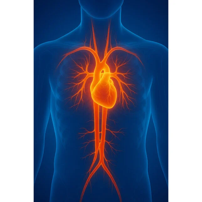
CT angiography of the chest (chest CTA) is a computed tomography scan taken while an iodine-based contrast agent flows through the blood vessels. The contrast is injected into a vein in the arm, and a technique called bolus tracking starts the scan as it reaches the vessels in question: the pulmonary arteries in a CT pulmonary angiogram (CTPA), or the thoracic aorta in a CT aortogram. Within seconds, the machine creates hundreds of thin slices that can be viewed in any plane or rebuilt in 3D.
A chest X-ray cannot show a clot inside an artery or a tear in the wall of the aorta. CTA can, without an invasive catheter procedure, which is why it has become the first-choice test for both. However, one study may contain more than a thousand images, and a small clot in a lower-lobe branch is easy to miss. In textbooks, an embolus is marked with a black arrow. On a real scan nothing points the way, so the radiologist has to follow every branch, from the main pulmonary artery to the edge of the lung.
How to order your CT angiography of the chest scan report
- Select your options. “CT angiography of the chest” is already chosen above. Add a translation or a comparison with an earlier examination if needed, then pay.
- Tell us why the scan was done. Note your symptoms, such as sudden breathlessness or chest pain, your history and your question. For a second opinion, attach the existing report.
- Send the images. You will then receive upload instructions. Hospitals usually release scans on a CD, DVD or USB storage device, or through a download link. We need the complete DICOM study, not screenshots.
- Specialist reading. A radiologist experienced in chest imaging reviews every series and, if you ordered a comparison, measures any change against your earlier scans.
- Download your report. We text you as soon as it is ready, typically two to three working days after your images arrive in full; five working days is our upper limit.
Why patients choose Eurodiagnosis
- Human expertise, signed by name. The radiologist whose electronic signature appears on your report is the doctor who examined your images and wrote every finding. Nothing in it is generated by AI or delegated to a trainee.
- Images before opinions. Your scan is judged on what it shows. An earlier report is useful background, but it is checked against the images rather than copied.
- Two readers, one document. The findings use the terminology your doctor works with, and the conclusion explains in everyday language what they mean for you as a patient.
- Secure. Your data is handled in line with GDPR.
What does a chest CT angiogram show? From the pulmonary arteries to the chest wall
A standard CT angiography report has two core parts: technical data and the assessment of the blood vessels. It opens with the technical data of the scanner, the way the contrast agent was administered (volume, injection rate, phase) and image quality. The radiologist then assesses every structure within the scanned area:
- Pulmonary arteries: analysed for contrast filling defects, dark gaps in the bright vessel that indicate pulmonary embolism. The search runs from the pulmonary trunk, which normally travels 4 to 5 cm before it divides, through the right and left branches down to segmental and, where image quality allows, subsegmental level. A widened main pulmonary artery can suggest pulmonary hypertension.
- The heart: the dimensions of the heart chambers, the right ventricle compared with the left (a ratio of 1.0 or more is a key sign that a clot is straining the heart) and fluid around the heart.
- Thoracic aorta: assessment covers the dimensions of the aorta and a search for aneurysm, as well as dissection, intramural haematoma, penetrating ulcer and atherosclerotic plaque.
- Main veins: the width of the superior and inferior vena cava.
- Lungs, airways and pleura: the trachea and bronchi, infarcts, consolidation, emphysema and fluid.
- Mediastinum, soft tissue of the chest wall and bones: lymph nodes, oesophagus, thyroid, ribs and spine.
- Upper abdomen: the parts of the liver, spleen and adrenal glands on the lowest slices. Contrast flowing back into the hepatic veins may be another sign of right heart strain.
Critical diameters are checked against reference values for the patient’s age and sex. For orientation, the right interlobar pulmonary artery is normally under 16 mm wide in men, the adult male trachea is 10 to 12 cm long and 13 to 27 mm across, and the left main bronchus is about 5 cm long. The report also covers extravascular structures, including incidental findings in the lung parenchyma, such as a small nodule, with a follow-up recommendation where needed.
How do I interpret a chest CT scan report?
Start with the conclusion, a short summary at the end. It answers the clinical question and flags life-threatening conditions first, such as an acute aortic syndrome or an embolus in the pulmonary trunk. Abnormal CT angiography results most often indicate pulmonary embolism, aortic pathology or atherosclerotic changes. When the findings could fit more than one condition, the report names the most probable first, then any others worth considering. A normal result is stated just as clearly: no abnormality detected, with no filling defects, an aorta of normal calibre, a heart of normal size and contour, and no enlarged lymph nodes. If breathing movement or poorly timed contrast limits the evaluation of some vessels, the report says so, because a technically limited study should not be read as a normal one.
Why would a doctor want a chest CT scan of the blood vessels?
In the UK, every CT exposure must be justified under the Ionising Radiation (Medical Exposure) Regulations (IR(ME)R). CT angiography of the chest is most commonly requested for:
- suspected pulmonary embolism: NICE recommends CTPA when a clot is likely on the two-level Wells score, or when it seems unlikely but a D-dimer blood test is positive
- sudden, tearing chest or back pain, to look for aortic dissection or other acute aortic disease
- monitoring a known thoracic aortic aneurysm, or checking the aorta after surgery or stent-graft repair
- unexplained breathlessness or suspected pulmonary hypertension
- chest trauma with possible injury to the aorta or other large vessels
- vascular abnormalities such as pulmonary arteriovenous malformations or obstruction of the superior vena cava
How is a CT angiogram of the chest performed?
The scan requires a small cannula in a vein in your arm. A radiographer then helps you lie on your back, usually with your arms raised above your head. A pump injects the contrast, typically at 4 to 6 ml per second, followed by a saline flush that keeps it in a compact bolus for sharper images. You hold your breath for a few seconds while the images are taken. Many patients notice warmth, a metallic taste or a feeling that they have passed urine (they have not), which fades within a minute or two. The scan itself takes only a few minutes, but allow 30 to 60 minutes in the department, including a short wait after the injection.
Contraindications and safety
Tell the CT team in advance about any of the following:
- Kidney disease or diabetes, or taking metformin. A recent eGFR blood test may be required.
- A previous reaction to contrast, or an overactive thyroid. Staff will decide on precautions or another test.
- Pregnancy or possible pregnancy. A clot in pregnancy is serious, so imaging is not withheld. The team may first scan your legs with ultrasound and will discuss CTPA or a lung perfusion scan with you.
- Breastfeeding. UK guidance does not require you to stop after a contrast injection.
The UK Health Security Agency (UKHSA) dose comparison table gives a typical CT scan of the chest as approximately 6.6 mSv, compared with 0.014 mSv for a chest X-ray and 2.6 mSv a year from all sources for an average person in the UK. A Eurodiagnosis report uses images you already have and therefore adds no radiation.
Clear answers about the vessels of your chest
Phrases such as “borderline aortic dilatation” can raise more questions than they answer. A specialist reading explains what such wording means and, where the images allow, gives a definite answer: what was found, where, how large it is and what should happen next. In addition, during aneurysm monitoring, the comparison option records any change in diameter.
Your chest CT angiogram, read by a radiologist: no referral, no waiting list, no clinic visit.
CT angiography of the chest: frequently asked questions
No. The only discomfort is the cannula, much like a blood test. The warm feeling from the contrast is common, normal and short-lived.
For a planned scan, usually yes, because no sedation is given. If you received medicine to help you relax or you feel unwell, arrange a lift. If you are being assessed for a suspected clot as an emergency, do not drive yourself.
Most patients have it within three working days of a complete upload. An urgent finding is stated at the very top of the conclusion. If you have not already been treated for it, contact your doctor or call 999 straight away.
No, you can order directly. Adding the reason for the scan, or the original request, lets the radiologist answer your doctor’s exact question.
Yes, whether it was done by an NHS trust, a private clinic or a hospital abroad, as long as you send the original DICOM files.
Yes, both are covered here. Add the type of scan to your order notes if you know it. If the study also includes the abdomen and pelvis, call our freephone helpline first.
Doctors generally welcome a structured, electronically signed specialist report, although how they use it is up to your GP or consultant.