White spots are among the findings radiologists note most often on a brain MRI, and they regularly appear in people who feel perfectly well and were scanned for an unrelated reason. That very commonness is where the confusion starts: a white spot can reflect anything from an ordinary, age-related change to a condition that warrants follow-up, so on its own it settles very little. What gives it meaning is the wider context – its location, its appearance, the person’s age and medical history, and how the full study reads to a specialist. Understanding roughly what these areas are, and why they are never read in isolation, can make the wait for a report considerably less stressful.
White matter, gray matter, and where white spots come from
The brain is built from two main types of tissue: gray matter and white matter. White matter takes its name from the fatty material called myelin that coats the nerve fibers running through it, and this insulation is what keeps signals moving quickly enough to support normal brain function across the central nervous system. White spots almost always sit within this white matter, which is precisely why they are so often labelled white matter lesions.
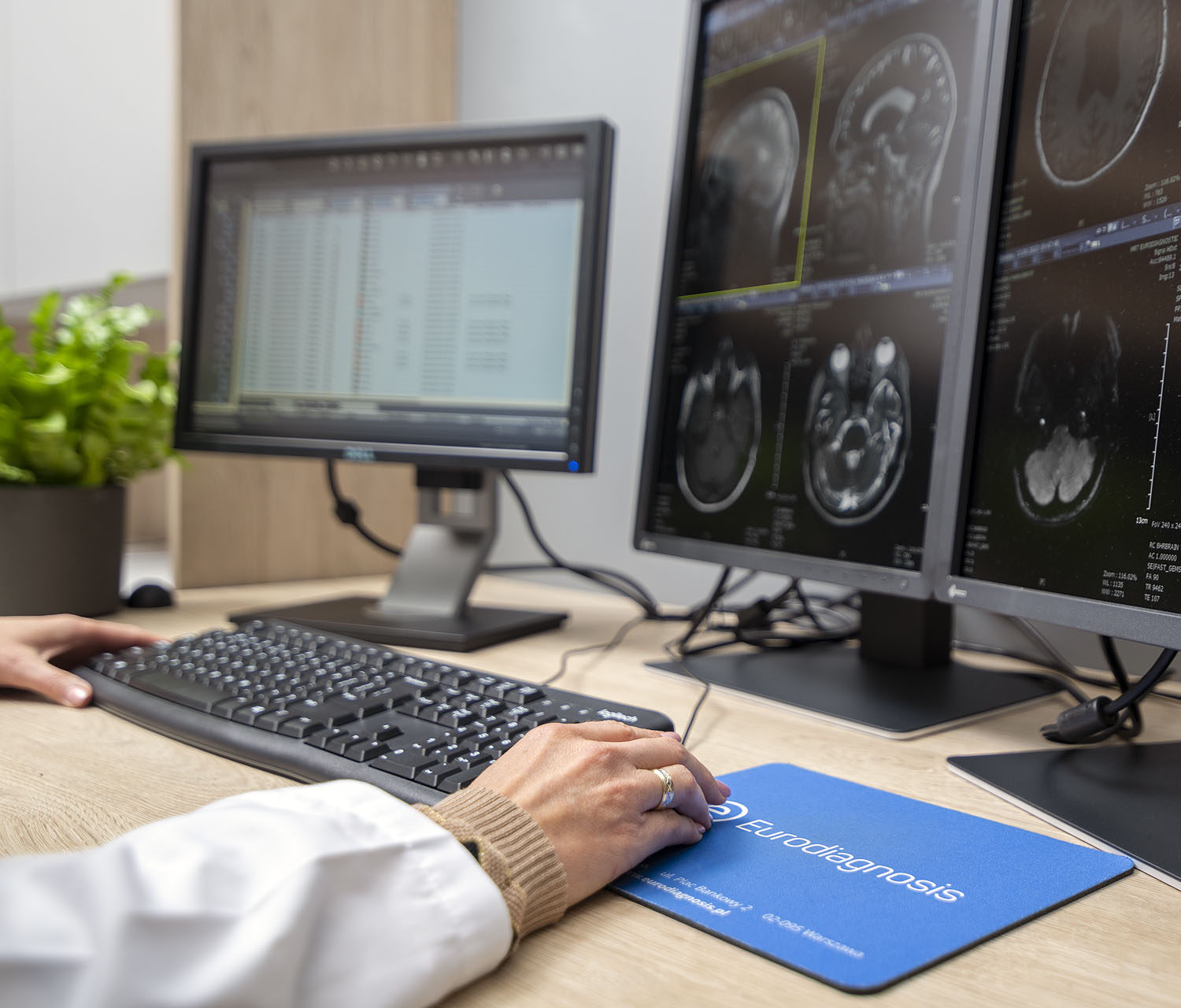
Every tissue gives off a measurable signal during a brain MRI, and the strength of that signal is what produces the bright areas and darker areas you see on the image. Healthy brain tissues return a fairly even pattern; when myelin, nerve fibers, or the surrounding brain cells are disturbed, that small patch sends back a different signal and can form lesions that stand out against the background. What a non-specialist reads as a white spot is, to a radiologist, simply a localized change in signal intensity.
How obvious these areas look is not fixed. It depends on which MRI sequences are run, on the imaging techniques chosen for that examination, and on the region being studied – the same patch can appear prominent on one sequence and almost invisible on another. Because magnetic resonance imaging relies on a magnetic field and radio waves rather than ionizing radiation, the same area can be revisited and re-imaged from several angles without the concerns that come with other methods.
White spots on a brain MRI: what does it mean?
White spots are often reported as white matter lesions or, more broadly, brain lesions. They are among the most common incidental findings on brain scans, meaning they are frequently noticed during imaging done for an unrelated reason. Their presence is a starting point for assessment, not a conclusion. The same finding can carry different significance in two different people, which is why a single image is never enough to decide whether a disease is present. Where the area sits also matters: signals along the brain and spinal cord are read differently from one another, and the spinal cord itself is assessed with its own considerations.
Common causes of white matter lesions
There are many possible explanations, ranging from harmless to those that need follow-up. Depending on their number, size, and location in different parts of the brain, white spots may be linked to:
- Small vessel disease and vascular changes. This is one of the most frequent causes, particularly later in life. When small blood vessels narrow and blood flow is reduced, areas of the brain’s white matter can be affected and form lesions. High blood pressure, high cholesterol, heart disease, and other vascular risk factors raise the likelihood of these changes, and people with several cardiovascular risk factors may be at higher risk.
- Normal aging. As people age, some white spots simply reflect normal aging of the small blood vessels and surrounding tissue. Research shows that white matter lesions become more common in older adults, and many of these areas cause no noticeable symptoms.
- Inflammatory and demyelinating conditions. Multiple sclerosis is a condition of the central nervous system in which the protective myelin around nerve fibers is affected. It can appear in younger people and is one reason inflammatory conditions are considered when lesions follow certain patterns.
- Other factors. Migraines, vitamin deficiencies, exposure to certain toxic substances, and some infections can also be associated with changes in the white matter. Identifying the underlying cause depends on the full clinical picture, not on the scan alone.
Are normal white spots on a brain MRI a cause for concern?
Not every finding signals a problem. Small white spots on a brain MRI are often incidental, especially in older adults, and many people who have them experience no noticeable problems in daily life. Whether white spots are considered normal depends on several factors: how many there are, their size and shape, their location, the person’s age, and whether there are any symptoms. A small number of stable spots is interpreted very differently from a pattern of new lesions or more lesions appearing over time. Because of this, normal white spots on a brain MRI and findings that need attention can look superficially similar, and only a specialist can tell them apart in context.
Symptoms sometimes linked to white matter lesions
In many cases, white matter lesions are found without the person having any related complaints. When symptoms do occur, they depend on the cause and on which brain cells and nerve fibers are involved. Reported symptoms can include memory loss or other signs of cognitive decline, balance problems, muscle weakness, vision problems, and mood changes. These are not specific to one diagnosis, and significant damage is usually associated with more advanced or severe cases rather than with small, isolated findings. Anyone experiencing such symptoms should discuss them with a healthcare provider rather than drawing conclusions from the images.
How specialists interpret the findings
A white spot on its own tells a radiologist very little; what matters is the company it keeps. The assessment weighs how many spots there are, their size and shape, exactly where they sit in the brain or along the spinal cord, and whether the overall pattern is typical of vascular change, demyelination, or something else. Comparing the current images with any earlier brain scans is often decisive, because a finding that has stayed unchanged for years is read very differently from one that has just appeared. Where more information is needed, a contrast agent may be given, since the way a region takes up contrast can help separate active processes from old, settled ones. As some contrast agents are cleared by the kidneys, simple blood tests are occasionally done first to confirm kidney function before the dose is given.
Not every bright patch is a true lesion, and part of the work is ruling out look-alikes. Movement during the scan can blur the image and create false impressions, and metal inside the body can do the same by distorting the magnetic field around it. Surgical clips, spinal or orthopedic fixation, and dental work are common sources of this, while a few implanted devices – a cochlear implant being the classic example – rule the examination out altogether for safety reasons. This is why staff ask, before a brain MRI, about any implants, retained metal, earlier operations, current medications, and the possibility of pregnancy: the answers protect both the safety of the examination and the reliability of the images. The scan itself is non-invasive.
Once these factors are accounted for, the radiologist records the size, shape, and character of any lesions in the MRI report and sets them against the rest of the study. That report is a description rather than the final word: the referring physician, and a neurologist where the picture points that way, weigh the MRI results together with symptoms and history before deciding whether anything needs to be done. If the underlying cause remains uncertain, further imaging or other tests may follow.
Addressing the underlying cause
Because white spots are not a single condition, there is no single response to them. Treatment options depend entirely on what is driving the findings. Where vascular disease or small vessel disease is involved, management often focuses on the contributing risk factors: monitoring blood pressure, managing high cholesterol, quitting smoking, and addressing a poor diet through lifestyle changes that support overall brain health. In inflammatory conditions, the aim may be to reduce inflammation and treat the specific neurological conditions identified. Physical therapy and other supportive measures can help with certain symptoms. In situations where stability matters, follow-up imaging is sometimes used to check whether new lesions have developed. Decisions about all of this rest with the treating physician.
Remote MRI description with Eurodiagnosis
If you already have images from a brain MRI but no accompanying written report, Eurodiagnosis provides a remote reporting service for MRI scans. You send the images online through a secure system, a radiologist experienced in interpreting magnetic resonance imaging reviews them, and you receive an electronically signed report, typically within a few working days. The report is an interpretation of the submitted images; decisions about any further diagnostics and treatment remain with the referring physician and the treating doctor. Details are available on the MRI scan report page, and an overview of the full service on the request a report page.
Frequently asked questions
What does it mean if my MRI report mentions white spots?
It means the radiologist noticed one or more areas in the white matter whose signal differs from the surrounding brain tissues. On its own this does not indicate a specific diagnosis; the meaning depends on the number, size, location, and pattern of the findings, as well as your age and symptoms.
Are white matter lesions always serious?
No. Many are incidental and stable, particularly in older adults, and are linked to normal aging or vascular changes. Others may point to conditions that need monitoring or treatment, which is why interpretation is left to a specialist.
Do white spots on a brain MRI mean multiple sclerosis?
Not necessarily. Multiple sclerosis is one of several possible causes, and it tends to follow particular patterns in the central nervous system. Vascular changes and aging are far more frequent explanations, especially as people age.